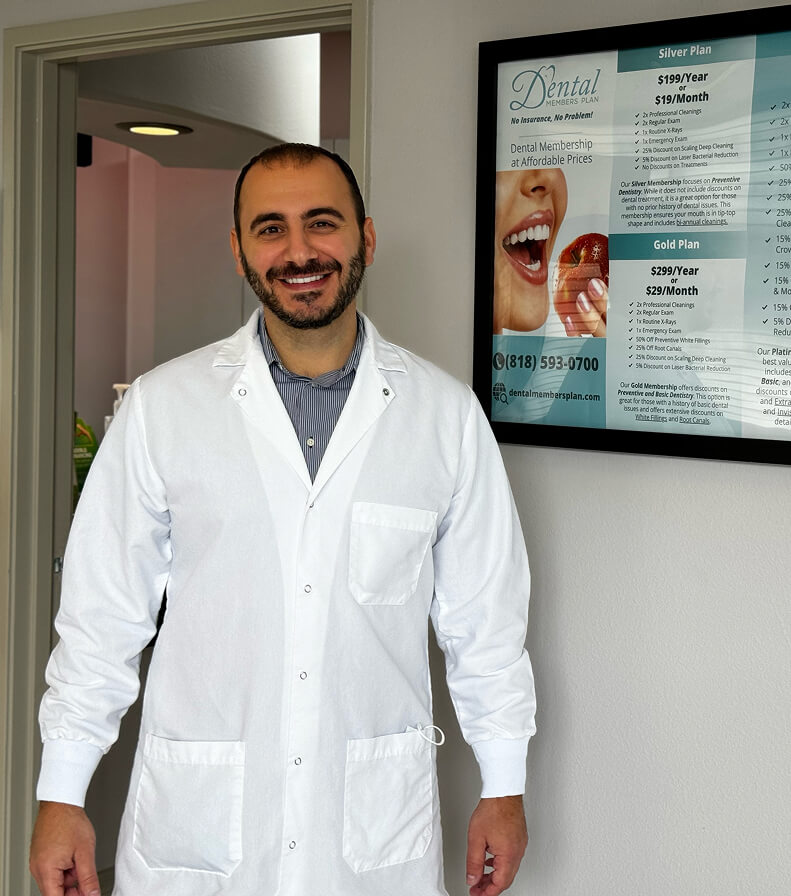
A-Dental Center has been a trusted part of the North Hollywood community since 1998. Dr. Fadi Elzayat has led the practice since 2008, bringing over 20 years of clinical experience in dental implants, All-on-4, full arch restorations, and comprehensive family dentistry.
A graduate of the USC Herman Ostrow School of Dentistry, one of the top dental schools in the country, Dr. Elzayat holds a Fellowship from the International Congress of Oral Implantologists (FICOI) — a distinction held by fewer than 5% of implant dentists worldwide, making it one of the most prestigious credentials in the field of implant dentistry.
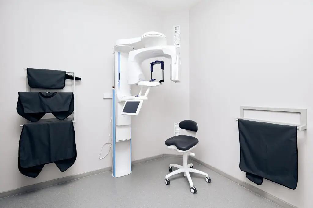
He focuses on cosmetic dentistry, with a strong emphasis on dental implants and advanced full-arch solutions. He performs every implant case personally and in-house, often utilizing Cone Beam 3D imaging for precision planning.
No referrals. No outside oral surgeons. Every phase of your treatment, from consultation to final restoration, is handled or supervised by Dr. Elzayat at A-Dental Center.
Beyond clinical skill, Dr. Elzayat is known throughout North Hollywood as “The Friendly Dentist” for his calm, straightforward approach, gentle technique, and zero-pressure consultations. A strong focus on comfort and clear communication ensures that each visit feels personalized and supportive.
Whether you haven’t seen a dentist in years or are facing a complex full arch case, patients from North Hollywood, Burbank, Studio City, and the greater San Fernando Valley are welcomed into a setting designed for a relaxed, judgment-free experience.
Credentials:
 USC School of Dentistry Graduate
FICOI Fellow, International Congress of Oral Implantologists
20+ Years Placing Dental Implants
Bilingual, English & Spanish team
ADA, CDA & San Fernando Valley Dental Society Member
USC School of Dentistry Graduate
FICOI Fellow, International Congress of Oral Implantologists
20+ Years Placing Dental Implants
Bilingual, English & Spanish team
ADA, CDA & San Fernando Valley Dental Society Member
 USC School of Dentistry Graduate
 FICOI Fellow, International Congress of Oral Implantologists
 20+ Years Placing Dental Implants
 Bilingual, English & Spanish team
 ADA, CDA & San Fernando Valley Dental Society Member